Contacts : nikolay.cherkashin@cemes.fr, lionel.calmels@cemes.fr, remi.arras@cemes.fr
GeSbTe (GST)-based alloys are phase-change materials, meaning they exhibit vastly different physical properties depending on whether they are in an amorphous or crystalline state. In DVDs and Blu-ray discs, binary information is stored using the sharp contrast in optical reflectivity between these two phases.
In electronic phase-change memory (e-PCM), information is encoded in the marked difference in electrical conductivity between the crystalline and amorphous phases of these alloys. These states are then switched locally and reversibly between the crystalline and amorphous phases using thermal pulses generated from electrical pulses. Heating above the melting temperature followed by rapid quenching allows the formation of an amorphous region with high resistivity, whereas more limited but prolonged heating allows the recrystallization of this same region.
Recent work, including our own, has demonstrated that beyond digital memories (2 bits), these alloys can also be used to fabricate multilevel memories (multiple bits) as well as memristors, capable of replicating synaptic activity, and thus offer devices for artificial intelligence.
Despite their enormous potential, the development and industrialization of e-PCMs require a thorough understanding of the physical phenomena involved in switching and storage mechanisms, particularly at extremely small scales. Currently, most integrated circuit manufacturers are exploring the potential of these materials in collaboration with academics, and this project is no exception.
A simplified diagram of the architecture of an e-PCM cell, along with a TEM image, is shown below. It consists of a heating filament (TiSiN), the phase-change material (GST), and the top electrode (TiN). The TEM image shows the cell’s characteristic amorphous dome in the RESET state (0). The dimensions illustrate the need to conduct studies at the nanoscale using advanced microscopy techniques.


On the left, a simplified diagram of the “mushroom” structure of PCM cells. On the right, an SEM image of an actual cell in the RESET state. Note the amorphous dome above the heating filament.
Contact : nikolay.cherkashin@cemes.fr
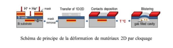
The selective manipulation of the electronic structure of new 2D materials through deformation opens up new opportunities in nanoelectronics, particularly in high-frequency (HF) transistors. To take advantage of the high electron mobility in graphene, it is necessary to create a bandgap; this could be achieved by applying enormous stress perpendicular to the graphene plane, using a “bottom-up” approach based on the growth of blisters beneath the graphene.
Contact : nikolay.cherkashin@cemes.fr
We are conducting research to identify the mechanisms and effects induced during ion implantation using the Smart-Cut™ process. This research is being carried out in collaboration with SOITEC.
A notable example of the application of this approach involves the ion implantation of H+ and He+ into Si. When followed by molecular bonding, this process can be used to fracture and transfer thin Si layers from a donor substrate to a host substrate, providing the most efficient method (Smart-Cut™ technology) for the fabrication of SOI (silicon-on-insulator) substrates—a field in which France, through SOITEC, is a world leader. The fracturing of Si results from the growth of “platelets”—flat cavities filled with molecular hydrogen—and their elastic interactions, which lead to the formation and growth of nano- and microcracks or pressurized blisters (Figure below).

Defects, precipitation, maturation, coalescence, transformation, and fracture following H/He implantation in Si: a multiscale study.
In fact, all of these systems (matrix/precipitates) result from the precipitation, during thermal annealing, of high supersaturations of point defects (chemical impurities, interstitials, and vacancies) initially introduced into the matrix by ion implantation. These defects altered the lattice volume of the host matrix and therefore were expected to induce biaxial stress in the matrix plane [1] (see next figure).

(a) MET cross-sectional image; deformation (b), (c) out-of-plane εzz; (d), (e) in-plane εxx obtained in Si implanted with H using (b), (d) dark-field holography, (c), (e) FEM using the sub-nanometer defect model (f). The image on the right shows the material’s elastic response to the presence of the complexes and the stress σxx.
Consequently, this constraint has a significant impact on the type (structure, families, variants), growth rate, and any transformations of the complexes [2], extended defects, and precipitates that form during the thermal annealing of these supersaturated matrices.

Profiles of the various complexes (a) and the contribution of the various complexes to macroscopic deformation (b), (c), derived for two doses of implanted hydrogen.
Since these characteristics, which are experimentally measurable, directly reflect the formation energy of the precipitates, our idea was to generate and use anisotropic fields to influence these populations and thereby access all components of the Gibbs energy of 2D precipitates. This approach allowed us to trace back to the fundamental parameters of precipitation—formation enthalpies and bond energies of the species, the elastic, surface, and volume energies involved, and the diffusivities of the species—and ultimately to simulate the phenomenon [3].
[1] N. Cherkashin, S. Reboh, A. Lubk, M. J. Hÿtch, A. Claverie, “Strain in Hydrogen-Implanted Si Investigated Using Dark-Field Electron Holography”, Applied Physics Express 6, 091301 (2013).
[2] N. Cherkashin, F.-X. Darras, P. Pochet, S. Reboh, N. Ratel-Ramond, A. Claverie, “Modelling of point defect complex formation and its application to H+ ion implanted silicon”, Acta Materialia 99 (15), 187-195 (2015).
[3] N. Cherkashin, F. X. Darras, A. Claverie, “Determination of the Free Gibbs Energy of Plate-Like Precipitates of Hydrogen Molecules and Silicon Vacancies Formed after H+ Ion Implantation into Silicon and Annealing”, Solid State Phenomena 242, 190-195 (2015).
Contact : remi.arras@cemes.fr
We are studying the properties of new ferroelectric materials compatible with CMOS technologies. For example, we calculate the formation of point defects near the boundaries of ferroelectric domains in ScxAl1-xN nitrides. These materials will subsequently be used to create interfaces of interest for spintronic applications (see section Materials for spinorbitronics and orbitronics) [Pezo 2024].
Contact: nikolay.cherkashin@cemes.fr
In general, the epitaxy of heterostructures requires the accommodation of materials with different crystal lattices. Since the substrate is volumetric and rigid relative to the deposited layer, the lattice of the deposited layer deforms and generates biaxial stress in the substrate plane. Consequently, this layer stores elastic energy anisotropically, which significantly impacts the behavior of the system during growth or when a second phase is precipitated within it. A well-studied example in the scientific community involves the growth of pseudomorphic layers on a substrate or layers that are plastically relaxed through the formation of dislocations. Another example involves the transformation of a thin pseudomorphic layer (2D) into separate islands (3D) due to the relaxation of the elastic energy accumulated in the 2D layer. We set out to develop experimental and theoretical methods to describe elastic and plastic relaxation phenomena under stress in III-V materials. We have thus developed a relatively simple analytical 3D model that allows us to deduce, from strain images obtained by MET (GPA on HREM), the local stoichiometry of InGaAs islands in a GaAs matrix and thereby to understand, simulate, and control the optical properties of lasers using these quantum dots [1, 2].

Out-of-plane deformation (HR-DFEH), elastic deformation (FEM), localization potentials, and hole wave functions (calculation) in an InGaAs/GaAs quantum well.
[1] 1. N. Cherkashin, S. Reboh, M. J. Hÿtch, A. Claverie, V. V. Preobrazhenskii, M. A. Putyato, B. R. Semyagin, V. V. Chaldyshev, “Determination of stress, strain, and elemental distribution within In(Ga)As quantum dots embedded in GaAs using advanced transmission electron microscopy”, Applied Physics Letters , 102:173115 (2013).
[2] A. N. Kosarev, V. V. Chaldyshev, and N. Cherkashin, Experimentally-Verified Modeling of InGaAs Quantum Dots, Nanomaterials, MDPI, 12 (12), 1967 (2022).
[3] A. F. Tsatsulnikov , W. V. Lundin , A. V. Sakharov , A. E. Nikolaev , E. E. Zavarin , S. O. Usov , M. A. Yagovkina , M. J. Hÿtch , M. Korytov, and N. Cherkashin, “ Formation of Three-Dimensional Islands in the Active Region of InGaN Based Light Emitting Diodes Using a Growth Interruption Approach”, Science of Advanced Materials 7(8):1629-1635 (2015).
Contacts : remi.arras@cemes.fr, lionel.calmels@cemes.fr
Contacts : dongzhe.li@cemes.fr, anne.bernand-mantel@cemes.fr
contact : nicolas.biziere@cemes.fr
For several years, our group has been developing a new methodology based on the principle of electron holography in a transmission microscope to image spin wave modes in magnetic nano-objects at the nanometer scale. One of the advantages of this new approach is that it allows us to correlate the spatial profile of the modes with the equilibrium magnetic configuration and the geometric and/or chemical imperfections of the object. In particular, this method will help improve theoretical and numerical models describing dynamic modes in highly inhomogeneous equilibrium magnetic configurations. To achieve this goal, we have developed two sample holders that allow the injection of electrical signals up to 18 GHz into an electron microscope; one features two microstrip-type electrical channels, and the other four. This technological achievement, unique on an international scale, is the result of a collaboration with the companies CISTEME and A3D design. This has made it possible, for the first time, to observe via electron microscopy the precession of the uniform mode at the center of a permalloy nanoribbon, as well as the complexity of the precession mode of the double-vortex closure domain.
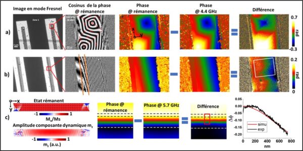 Fresnel-mode images and phase images in holography for the remanent state and the excited state at 4.4 GHz for the line head (a) and the line center (b). (c) Micromagnetic simulations of a 10 µm × 1 µm × 40 nm line showing the remanent state and the quasi-uniform mode profile at 5.7 GHz in the simulation (left side), and the corresponding reconstructed phase images. The figure on the right shows the experimental and simulated variations in phase amplitude along the Y direction, averaged over the surfaces bounded by the white and red squares.
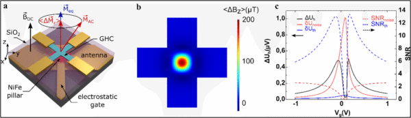 a) Principle of FMR mode detection in a permalloy patch with a graphene Hall cross. b) Variation of the out-of-plane component of the time-averaged radiated field at 30 nm above the patch between the static state (without RF excitation) and the dynamic state (with RF). The patch measures 500 × 500 × 20 nm, and the Hall cross has a diameter of 400 nm at its center. It can be observed that only the center of the patch oscillates strongly, leading to a highly localized variation in ∆Bz. c) Calculated sensor response (∆Uh) based on b). The solid-line curves correspond to the calculated total electronic noise (red) and thermal noise (blue) of the sensor. The dashed-line curves correspond to the calculated signal-to-noise ratio.
[1] Concept of imaging spin waves in pseudo-static mode by electron holography, Marine Resano, Christophe Gatel, Abraao Cefas Torres-Dias, Benjamin Lassagne and Nicolas Biziere, Appl. Phys. Lett. 127, 212404 (2025).
contact : nicolas.biziere@cemes.fr
X2YZ-type Heusler alloys are particularly promising for the fabrication of radiofrequency components (> 1 GHz) due to their low damping coefficient and magnetization value exceeding 1 T. Furthermore, these materials are potentially semimetallic with spin polarization on the order of 100%, which is important for designing giant magnetoresistance or tunneling sensors. Our team has long studied the influence of different types of crystalline disorder on the values of these magnetic parameters using broadband ferromagnetic resonance measurements (30 GHz). One of our current research activities involves leveraging these properties to develop reconfigurable radiofrequency components. For example, the absorption spectrum of a magnonic crystal based on square antidots can be significantly modified in the remanent state due to the material’s cubic anisotropy [1]. Indeed, under the influence of a weak, ultrashort field pulse (a few mT, 1 ns), it is possible to obtain remanent states that may or may not couple with a radiofrequency field [Fig. 1(b,c)]. Our future work in this area will focus on integrating these magnonic crystals into spin waveguides to realize zero-field frequency filter functions. To this end, we are specifically studying the impact of nanomanufacturing methods (FIB, optical and electronic lithography, Ar+ ion beam etching, etc.) [2] on the number of dynamic modes accessible in the system (Fig. 2).

Figure 1: a) SEM image of a magnonic crystal consisting of an array of square antidots with a side length of 300 nm and a spacing of 900 nm. The crystal was fabricated by electron lithography followed by etching with a 3 KeV Ar+ ion beam. b) Micromagnetic simulations of the remanent states A1 and A4 corresponding to two directions of the saturation magnetic field pulse. c) RF absorption spectra measured in the crystal for configurations A1 and A4, and micromagnetic simulations of the dynamic mode frequencies (blue) in configuration A4.

Figure 2: Evolution of absorption frequencies as a function of the external magnetic field in a magnonic crystal composed of a lattice of square antidotes with 100 nm sides, spaced 300 nm apart. The crystal is fabricated by Ga+ ion etching at 30 KeV using a FIB. On the right, an example of the spatial profiles of the dynamic modes obtained from micromagnetic simulations (taken from [2]).
[1] Cubic Anisotropy for a Reconfigurable Magnonic Crystal Based on Co2MnSi Heusler Alloy, S. Mantion and N. Biziere, Phys. Rev. Applied 17, 044054, (2022).
[2] Influence of Ga+ milling on the spin waves modes in a Co2MnSi Heusler magnonic crystal, S. Mantion and N. Biziere, J. Appl. Phys. 131, 113905, (2022).
Contact : nikolay.cherkashin@cemes.fr
For the study of nanocrystals embedded in amorphous matrices and epitaxial layers with very different crystal structures, we have been looking for a solution to measure the deformation from a high resolution image (HR-TEM or scanning probe TEM=HR-STEM) in the absence of a reference on the same image and/or the presence of random distortions. This is a problem we were facing, and is an always annoying constraint inherent to all other methods. To solve this problem, we proposed and developed a new method called “Absolute strain” (AbStrain) for HR-(S)TEM image processing allowing the measurement of absolute values of interplanar distances and angles in single crystal structures without the need to use a reference grating present on the same image. To this end, we developed a theory of “absolute correction” (as opposed to relative correction) of instrumental distortions affecting experimentally measured images and wrote a script to apply this theory to analyse nanoscale structures for which a reference grating could not be imaged. This approach can be applied not only in reciprocal space [1, 2]] (Fig. 6.1) (where the GPA technique operates) but also in real space (where the so-called peak findings technique operates).

Figure 6.1 AbStrain: (a) HR-TEM image of a SbxAs1-x nanoinclusion (hexagonal structure) embedded in GaAs (zinc-blend structure); (b)-(d) zero components of the strain tensor obtained by AbStrain with respect to an ideal hcp lattice of As0.1Sb0.9.
[1] N. A. Bert, V. V. Chaldyshev, N. A. Cherkashin, V. N. Nevedomskiy, V. V. Preobrazhenskii, M. A. Putyato, B. R. Semyagin, V. I. Ushanov, M. A. Yagovkina, “Metallic AsSb nanoinclusions strongly enriched by Sb in AlGaAsSb metamaterial”, J. of Appl. Phys. 125, 145106 (2019).
[2] M. A. Luong, N. Cherkashin, B. Pécassou, Ch. Sabbione, F. Mazen, A. Claverie, Effect of Nitrogen Doping on the Crystallization Kinetics of Ge2Sb2Te5, Nanomaterials, MDPI, 11 (7), 1729 (2021).
Contact : nikolay.cherkashin@cemes.fr
We have sought to develop a method to quantify micrometer-scale deformation with nanoscale spatial resolution and ~10-4 X-ray accuracy in epitaxial heterostructures composed of multiple multilayers, i.e., deeply implanted ionic regions. For this purpose, we have invented a technique called “Moiré by Sample Design” (MoSD). This method allows the measurement of deformations in single-crystal structures, in cross-section and in plan view, via the formation of “controlled” moiré fringes while using conventional microscopes. The idea is to make a sample in the form of a stack containing the structure to be measured and a known reference crystal. Images then reveal moiré fringes from which the deformation fields in the structure under study can be extracted. By adjusting the angle of rotation between the two blades, the period of these fringes can be adjusted and thus maps of the strain fields can be obtained with nanometer resolution [1, 2] (Fig. 6.2). Having developed a moiré image processing algorithm, MoSD demonstrates the possibility of mapping two-dimensional strain fields with nanoscale spatial resolution (≥ 2 nm), an ultimate accuracy of 1×10⁻⁴ and a field of view twenty times larger than offered by GPA; this technique can be implemented using a conventional transmission electron microscope of modest cost.

Figure 6.2 MoSD: In-plane exx and out-of-plane ezz strain measured by MoSD and calculated by FEM in: (a) Si1-xGex multilayers [110]: conventional image, moiré, and the profiles of exx and ezz; (b) 3D mechanically coupled In0.4Ga0.6P islands formed on (112)GaAs substrate; (c) 3D mechanically coupled SiGe islands formed on (001)Si substrate.
[1] N. Cherkashin, T. Denneulin, M. J. Hÿtch, “Electron microscopy by specimen design: application to strain measurements”, Scientific Reports 7, 12394 (2017).
[2] N. N. Ledentsov, V. A. Shchukin, Yu. M. Shernyakov, M. M. Kulagina, A. S. Payusov, N. Yu. Gordeev, M. V. Maximov, A. E. Zhukov, T. Denneulin, and N. Cherkashin, “Room-temperature yellow-orange (In,Ga,Al)P-GaP laser diodes grown on (n11) GaAs substrates,” Opt. Express 26 (11), 13985-13994 (2018).
Contact : dongzhe.li@cemes.fr
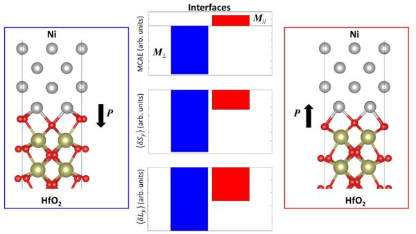
While early progress in spintronics relied mainly on phenomenological modelling (drift-diffusion and semi-classical Boltzmann transport equations), much more advanced quantum theoretical methodologies have made it possible to calculate magnetic and transport properties from realistic band structures obtained by ab initio methods based on density functional theory (DFT). However, due to the high computational cost, it is very difficult to model at the ab initio level magnetic skyrmions characterised by non-collinear spin textures of several nanometres width, induced by the spin-orbit coupling and resulting from the competition between different magnetic interactions. To this end, we will develop an ab initio large-scale spin-orbit transport platform that will allow us to model realistic quantum transport through skyrmions.
This CIFRE PhD is part of a collaboration between CEMES-CNRS (Toulouse) and Soitec (Bernin).
“Ô-GST” project:
Little is known about the physical and chemical changes that affect the material during phase transitions and that determine the cell’s electrical characteristics (resistive, conductive, or intermediate), or about the degradation mechanisms that affect it over time and with use. For this reason, fundamental research is needed to understand the mechanisms by which the material transitions from the amorphous phase to the crystalline phase (and vice versa), as well as the impact of the cell’s geometry, size, and surrounding environments on the final characteristics of the material and the associated device. Furthermore, the desired characteristics of the cells are achieved by using materials with clearly non-stoichiometric compositions, which further increases the need for an in-depth understanding of the atomic mechanisms involved and, consequently, for characterization at the nanoscale. In recent years, we have already explored crystallization and transport in Ge-rich GST alloys (GGST) as well as structure-property relationships in 28-nm technology cells and obtained some notable results that now define the state of the art in this field (1–10).

Phase separation during the crystallization of a Ge-rich GST alloy. Elemental mapping (MET-EDX) in layers annealed at 500°C and the corresponding ASTAR image (below). Large, pure grains of GST 225 are embedded in a matrix of fine-grained Ge.
In this context, CEMES is collaborating with STMicroelectronics on a major project called “Ô-GST,” whose objectives are to:
1) Identify the mechanisms and parameters governing the thermal crystallization of GGST materials and the changes resulting from doping with N, C, and H, both in deposited layers and within nanometric cells (28- and 18-nm technologies).
2) Understand the influence of GST domain morphology (phases, grain sizes, etc.) on the material’s electrical characteristics and on the performance and reliability (drift, retention/cycling) of PCMs based on these materials.
3) Explore the possibility of achieving intermediate resistivity states (IRS) and mimicking synaptic activity (analog storage, cumulative storage, and plasticity) using Ge-GST cells.
To achieve these objectives, we have formed a group consisting of three permanent researchers from CEMES and LPCNO with complementary expertise (theory, materials science, transport, and electrical properties), an expert engineer from STMicroelectronics, four postdocs, and a Ph.D. student.

A typical example of EELS chemical analysis of the active region of a PCM cell in the RESET state (resistive and amorphous). The forming step results in the creation of a typical nanostructure, consisting of Ge sidewalls and a GeTe roof, within which a homogeneous amorphous dome, rich in Ge and Sb, is formed. The total thickness of the layer is 50 nm.
This activity is strongly supported by direct contracts with STMicroelectronics (Task Force GST SOW1 and SOW2, Cifre) and by the European IPCEI projects on microelectronics implemented through the nano2022 and nano2025 programs, for a total amount of more than 2 million euros.

On the left, the evolution of cell resistivity as a function of the heat treatment applied. Initially, the amorphous material relaxes and becomes even more resistive (drift). Above 270°C, it becomes conductive.
Below, dark-field images show the stability of the Ge grains at the dome’s edge (in white), while GST-225 grains (in black) recrystallize and invade the dome starting at 260°C.