Using deep learning for segmentation in TEM
February 26, 2026
Is it possible to automatically extract quantitative and statistically significant parameters for studying dislocation dynamics? In this study, we take an important first step toward answering this question by demonstrating the possibility of using artificial intelligence to systematically detect dislocations under a wide range of transmission electron microscopy (TEM) observation conditions.
Dislocations, linear defects at nanoscale, are crucial in interpreting the mechanical properties of materials, but the quantitative study of their dynamics in TEM is time-consuming and potentially fragmented. The first obstacle to automating the analysis is the difficulty of detecting dislocations in the image.
The objective of the study published in Ultramicroscopy is to advance this task, known as segmentation, by determining for each pixel whether or not it belongs to a dislocation line. The difficulty here lies in the variety of dislocation contrasts generally observed in bright field/dark field, and the background noise inherent of the sample and microstructure.
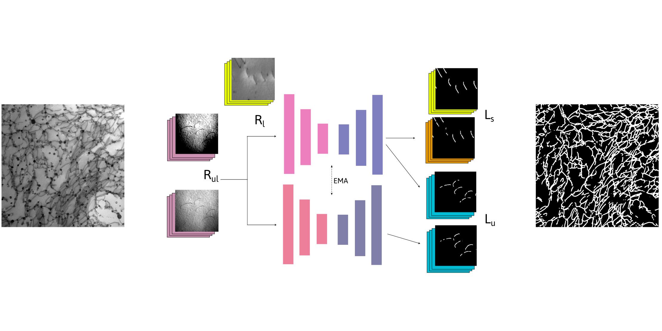
To solve this problem, we used several deep learning approaches based on neural networks. We began by training the network to segment a number of manually annotated images, a supervised approach. But we also had access to a large database of images and video sequences obtained in recent years by the PPM group at CEMES. We took advantage of this data by feeding it into a second network in parallel in order to improve its understanding of the intrinsic characteristics of the images. This semi-supervised approach significantly improves the model’s performance, bringing it much closer to that of “human” experts.
In order to completely eliminate the need for manually annotated images and expand the variety of observation conditions, we successfully created a database of synthetic images by combining Dislocation Dynamics and image simulations. Thanks to their high similarity, we show that these new images, while not improving the model’s performance on test images, do enable better predictions in more difficult situations that fall outside the usual TEM observation conditions.
Finally, we applied our model to the automatic calculation of dislocation densities in complex images of aluminum alloys. Our density estimates, made in a matter of seconds, are close to those made by hand… in over ten minutes!
Contact:
Frédéric Mompiou | frederic.mompiou[at]cemes.fr
Publication:
Deep learning approaches for dislocation segmentation in TEM
Assya Boughrara, Christine Viala, Laurent Dupuy, and Frédéric Mompiou
Ultramicroscopy 283 (2026) 114334
DOI: https://doi.org/10.1016/j.ultramic.2026.114334